

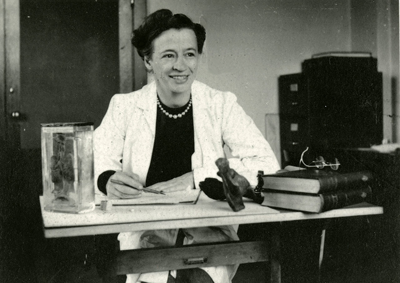
In November 1925, Maria Torrance Wishart founded the Department of Medical Art Service in the Anatomy Building (now the McMurrich Building) at the University of Toronto. It was the beginning of great things. Initially intended to supply medical images and models for U of T’s medical classes and teaching hospitals, the department was later expanded by Wishart to include a three-year diploma course. Today, it offers the university’s unique Master of Science in Biomedical Communications degree, one of just four accredited programs of its kind in the world.
But beyond being an incredible artist and dedicated teacher, Wishart also helped create space for women in the medical field, an area where they were marginalized for the first half of the 20th century.
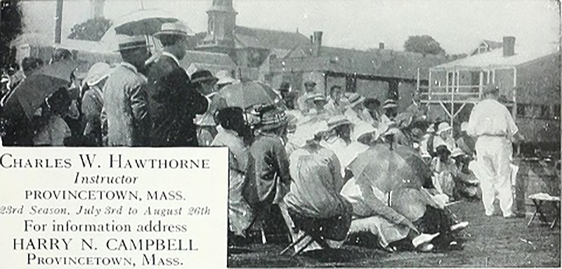
A young woman from Edwardian Toronto’s privileged class, Wishart traveled abroad and spent two years studying art and languages in Europe. When the First World War broke out in 1914, she returned to North America and took classes in anatomy, figure drawing and watercolour in Provincetown, Massachusetts.
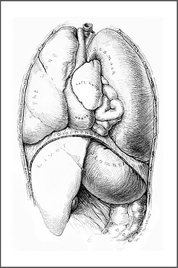
Most importantly for her future career, Wishart trained with the legendary medical illustrator Max Brödel, in Baltimore. In 1922, she had moved to Baltimore to study in the Art as Applied to Medicine Program (now in its 104th year) at Johns Hopkins University. There, she took classes in anatomy, physiology and pathology, along with applied art techniques suitable for reproduction (pen and ink, watercolour and carbon dust—a technique for which Brödel was famous).
Students remained in the program until Brödel felt they were ready to leave and a suitable position could be found for them. “Graduates” were sent off with a handshake instead of an official certificate or diploma. Wishart was a member of the 11-strong class of 1925, which remains the largest group of students to ever come out of the Hopkins program. Three quarters of the class were women and several of the students trained under Brödel, like Maria, went on to found medical illustration programs in the United States and Canada.
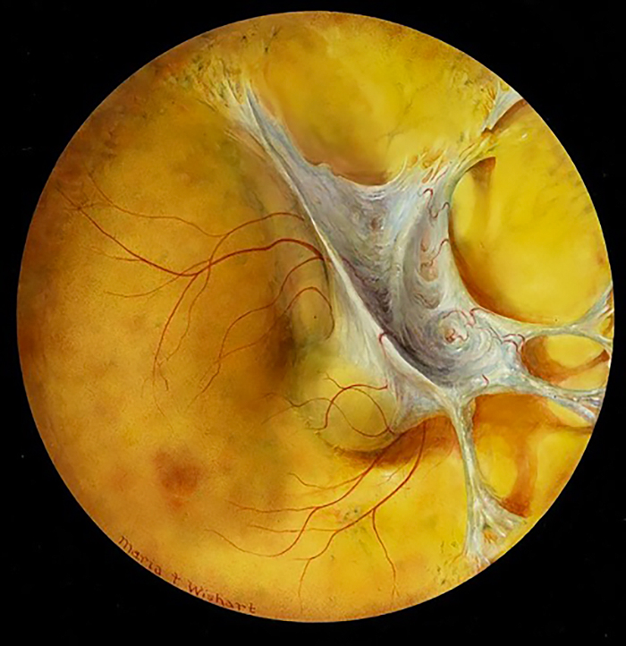
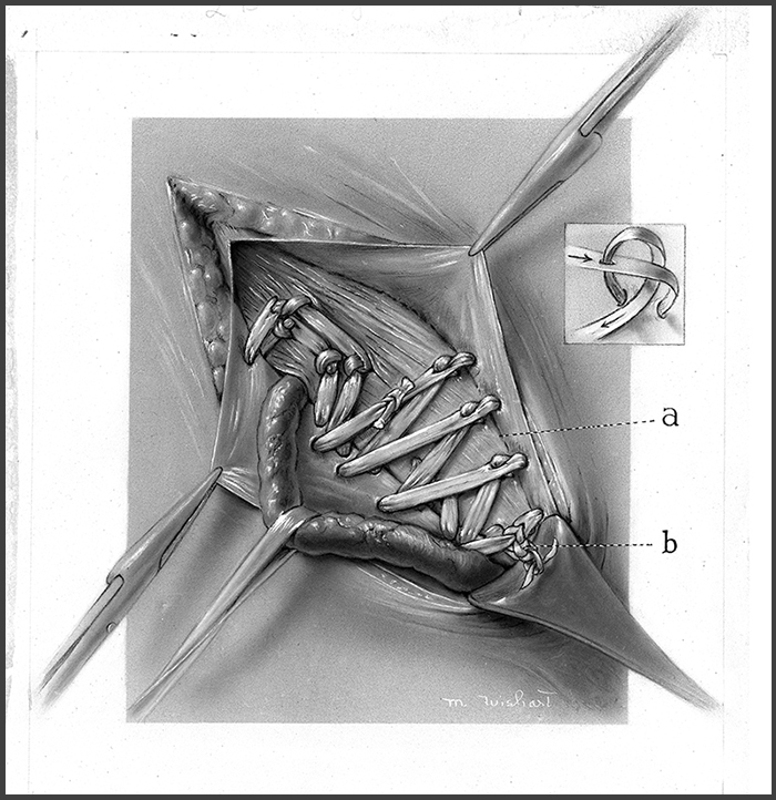
“Maria Wishart was an astoundingly talented and successful illustrator,” says Nicholas Woolridge, director of U of T’s Master of Science in Biomedical Communications Program. “She understood the importance of research, close observation, and careful and accurate representation. Good illustration is empathetic, in that it requires the illustrator to have a deep understanding of the needs of the viewer. Maria’s work demonstrated this quality by carefully emphasizing structure and its relation to function, eliminating unnecessary detail, and economically telling complex clinical stories. Her work was also exquisitely composed and rendered, and its beauty perhaps allows the clinical subject matter to be less distressing.”
In 1944, Wishart proposed an academic diploma program, Art as Applied to Medicine, to teach medical illustrators. The first students arrived in 1945 and the program mirrored the Johns Hopkins degree in admitting four women students for every man. As a teacher, Wishart adhered to the exacting standards set for her by Brödel. Students Victor Doray (Dip Art Applied to Medicine, 1956) and Marguerite Drummond (Dip Art Applied to Medicine 1946) (one of the first two graduates of the program) remembered her as, “dedicated, firm, compassionate, and memorable.”
Dedicated was right: she expected her students to work through lunch and on Saturdays to gain access to specimens when the medical students were away.
Wishart’s support of other career women was all the more extraordinary given her early background. She was born in 1893 into an affluent Toronto family. In the 1910s and ‘20s, her name appeared frequently in Toronto’s society pages, where, as a member of Toronto’s “young set,” her early life revolved around afternoon teas and evening dances. She spent summers in Georgian Bay and winters at skating parties and ski competitions. At age 19, she was among two dozen debutantes to “come out” in the 1912 season.
Wishart chose a path different from most of her debutante peers. And her artistic and teaching work was just the beginning. She went on to become a charter member and later president of the Association of Medical Illustrators. After retiring from the University of Toronto in 1962, she took classes at Central Tech in her later years and became an accomplished sculptor. She died in her ninetieth year in 1982.
One Response to “ Anatomy of an Illustrator ”
I recall my mother speaking of a Jean Wishart and also of a Wishart at U of T who did anatomical drawings for doctors. I wrote my sister for details and she replied, "This article is fascinating! Maria Wishart was Jean Wishart's sister (I think a younger sister but I'm not sure). Mum often spoke of her and her tremendous talent.
Jean was an artist and taught art at a school in Hamilton. Mum used to go to Go-Home Bay (Georgian Bay) with the Wisharts. The Go-Home Bay picture that hung on the wall over my sofa was painted by Jean at our island in 1956 -- a very late spring and the trees were just leafing out on the May 24 weekend. I also have a water colour done by their mother at Go-Home, probably when the girls were teenagers.
I believe their father was a doctor, and they had a brother, Staunton, who was also a doctor. Mum and I went to visit Jean at her little house in Hamilton, probably in the early 1980s; she was terribly bent with arthritis then.
Thanks for the memories!